Arthroscopy is a surgical procedure orthopedic surgeons use to visualize, diagnose, and treat problems inside a joint.
The word arthroscopy comes from two Greek words, “arthro” (joint) and “skopein” (to look). The term literally means “to look within the joint.”
In an arthroscopic examination, an orthopedic surgeon makes a small incision in the patient's skin and then inserts pencil-sized instruments that contain a small lens and lighting system to magnify and illuminate the structures inside the joint. Light is transmitted through fiber optics to the end of the arthroscope that is inserted into the joint.
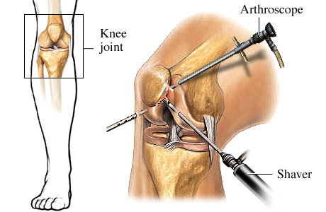
Diagnosing joint injuries and disease begins with a thorough medical history, physical examination, and usually X-rays. Additional tests such as magnetic resonance imaging (MRI) or computed tomography (CT) scan may be needed.
Through the arthroscope, a final diagnosis is made, which may be more accurate than through “open” surgery or from X-ray studies.
Disease and injuries can damage bones, cartilage, ligaments, muscles, and tendons. Some of the most frequent conditions found during arthroscopic examinations of joints are:
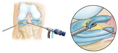
Although arthroscopic surgery has received a lot of public attention because it is used to treat well-known athletes, it is an extremely valuable tool for all orthopaedic patients and is generally easier on the patient than “open” surgery. Most patients have their arthroscopic surgery as outpatients and are home several hours after the surgery.